Pneumothorax
Pneumothorax, also known as a collapsed lung, occurs when air leaks into the pleural space—the area between the lung and chest wall. This air puts pressure on the lung, preventing it from expanding fully. It can cause sudden chest pain and shortness of breath, and in severe cases, it becomes life-threatening.
Types of Pneumothorax
- Primary Spontaneous Pneumothorax: Occurs without any known cause or underlying lung disease. It often affects tall, thin, young men.
- Secondary Spontaneous Pneumothorax: Happens in people with pre-existing lung conditions like COPD, asthma, or tuberculosis. It’s usually more serious due to poor lung reserve.
- Traumatic Pneumothorax: Results from blunt or penetrating chest injuries, including rib fractures or stab wounds. It may also occur during medical procedures like central line insertion or lung biopsy.
- Tension Pneumothorax: A medical emergency. Air enters the chest but cannot escape, increasing pressure and compressing vital organs. Immediate treatment is required.
Common Causes
- Lung disease (e.g., COPD, cystic fibrosis)
- Chest trauma
- Ruptured air blisters (blebs)
- Mechanical ventilation
- Sudden altitude or pressure changes
- Smoking (increases the risk significantly)
Symptoms of Pneumothorax
Symptoms may vary depending on the size and cause of the pneumothorax. Common signs include:
- Sudden sharp chest pain
- Shortness of breath
- Rapid heart rate
- Fatigue
- Bluish skin (in severe cases)
- Decreased breath sounds on one side
- Anxiety or restlessness
Large pneumothorax can cause low oxygen levels and life-threatening complications.
Diagnosing Pneumothorax
Physical Examination
Doctors listen to breath sounds and observe respiratory movements.
Imaging Tests
- Chest X-ray: Confirms air in the pleural space.
- CT Scan: Provides detailed images, especially useful in small or complex cases.
- Ultrasound: Quick and effective for bedside diagnosis in emergencies.
Treatment Options
Treatment depends on the size, severity, and cause of the pneumothorax.
- Observation: Small, stable cases may resolve on their own. The air is gradually absorbed by the body.
- Needle Aspiration: A needle is used to remove trapped air. It’s a common method for small to moderate pneumothorax.
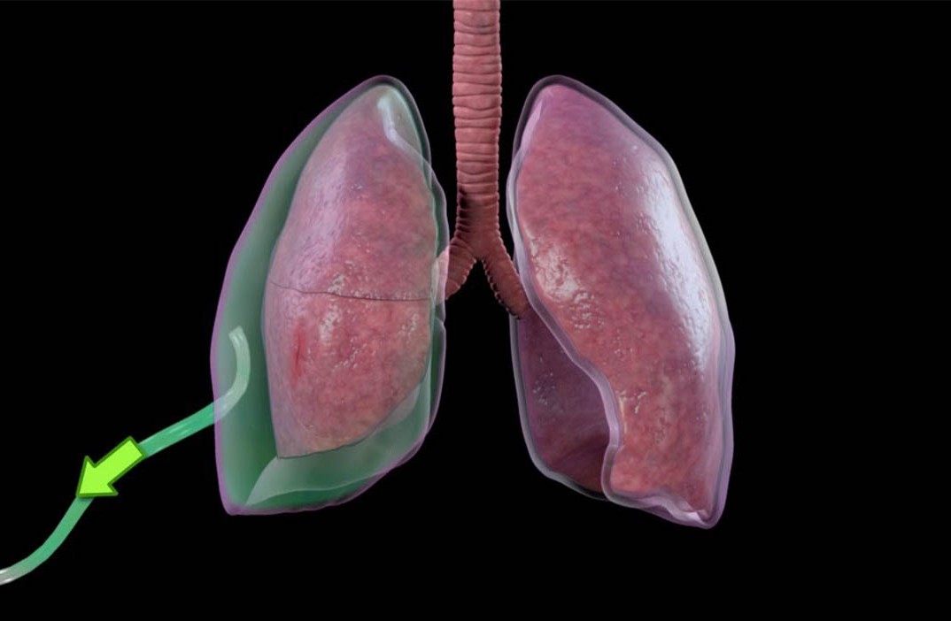
- Chest Tube Insertion: A tube is placed into the pleural space to continuously remove air. It stays in place until the lung fully re-expands.
- Surgery (VATS or Thoracotomy): In recurrent or large cases, surgery may be necessary to repair leaks and prevent recurrence. Surgeons may perform pleurodesis, which sticks the lung to the chest wall to stop future collapse.
Recovery and Aftercare
Most patients recover fully with proper treatment. Recovery time depends on the extent of lung damage and overall health. Avoid air travel and diving for several weeks after recovery. Smokers are strongly advised to quit to reduce recurrence risk.
Preventing Pneumothorax
- Don’t smoke.
- Manage chronic lung conditions effectively.
- Avoid extreme pressure environments if you're at risk.
- Get regular follow-ups if you’ve had pneumothorax before.
When to Seek Emergency Help
Call emergency services if you experience:
- Sudden chest pain
- Severe shortness of breath
- Fainting or confusion
These may indicate a life-threatening tension pneumothorax.